Ever caught a glimpse in the mirror and spotted those mysterious white patches staring back? You’re not alone. What starts as a minor curiosity can quickly become a nagging concern, quietly chipping away at your confidence and making you second-guess your comfort in your own skin. These seemingly innocent changes are rarely random; they’re your body’s way of signaling that something has disrupted the pigment-producing cells in your skin. The good news? While the triggers might be more common than you think, most causes are entirely manageable once correctly identified – yet, countless individuals delay seeking answers simply because these spots often don’t itch or hurt initially.
But here’s the crucial insight often missed by other articles: many of the most prevalent causes behind these white spots are linked to hidden, often modifiable triggers that are right under your nose. Unlocking this understanding isn’t just about treating symptoms; it fundamentally transforms how you care for your skin moving forward.
Unpacking the Science: What Truly Happens When White Spots Emerge
The vibrant, even tone of your skin is all thanks to melanin, a pigment meticulously crafted by specialized cells known as melanocytes. When these crucial cells are either diminished in quantity, suffer damage, or face a temporary blockage in their pigment-producing capabilities, lighter areas inevitably surface. The precise mechanism behind this varies significantly with the underlying cause – it could be a yeast byproduct disrupting pigment creation, your own immune system mistakenly targeting these cells, or even the cumulative effect of years of sun exposure gradually eroding them in localized patches.
Here’s the reassuring news: for the vast majority, these white spots are completely benign and pose no risk of contagion. The real challenge, and your path to effective management, lies in accurately identifying the specific type of spot you’re facing, allowing you to bypass guesswork and take precisely the right course of action.
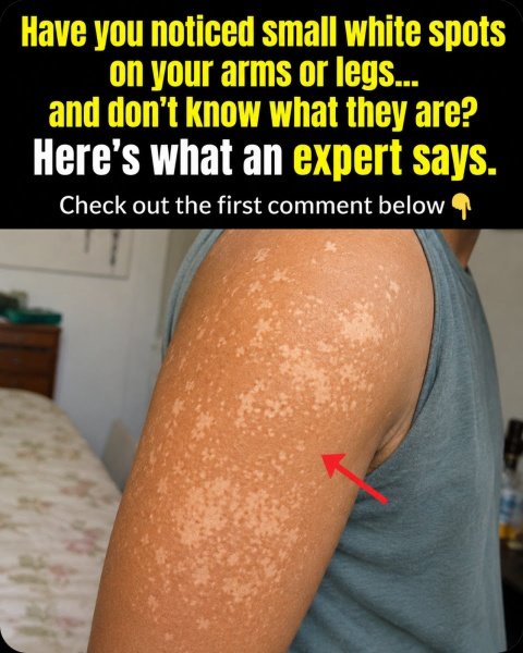
Tinea Versicolor: The Sneaky Yeast Overgrowth You Didn’t See Coming
Among the most common, yet frequently misidentified, culprits behind white spots is tinea versicolor (sometimes referred to as pityriasis versicolor). This isn’t an external infection in the traditional sense; rather, it’s an excessive proliferation of Malassezia yeast—a microorganism that naturally resides on everyone’s skin without causing issues under normal circumstances. However, when confronted with warm, humid environments or an increase in skin oiliness, this yeast secretes a specific substance that actively disrupts melanin production, resulting in the appearance of flat or subtly scaly patches that can manifest as white, pink, or even light brown.
You’ll typically find these distinctive patches manifesting on areas like the upper back, chest, shoulders, and upper arms. They tend to become strikingly more noticeable following sun exposure, primarily because the healthy skin around them develops a tan, while the compromised areas remain conspicuously lighter. It’s no surprise, then, that many individuals first spot them during the summer months or after returning from vacations in warmer, more humid regions.
What truly catches most patients off guard is learning that tinea versicolor bears no relation to poor hygiene; the Malassezia yeast is a permanent resident on your skin. The less obvious triggers include factors like profuse sweating, wearing restrictive synthetic fabrics that retain heat, recent antibiotic or steroid treatments, significant hormonal fluctuations, or even a subtle dip in immune system efficiency. While it’s neither dangerous nor readily transmissible, it does have a tendency to reappear if the environmental conditions remain conducive to its overgrowth.
Upon successful treatment with targeted antifungal shampoos or creams, the skin’s natural color typically begins to normalize gradually, a process that can span one to three months as your skin cells regenerate. The absolute critical step here is securing an accurate diagnosis; mistakenly applying potent steroid creams, for instance, can paradoxically exacerbate the condition and make the spots look worse.
Vitiligo: When Your Own Immune System Turns on Pigment Cells
Vitiligo is a condition where your body’s own immune system, in a perplexing error, launches an assault on its melanocytes—the very cells responsible for creating pigment. This leads to the characteristic appearance of smooth, distinctively milky-white patches, typically featuring well-defined borders. While it has the potential to manifest anywhere on the body, it frequently makes its first appearance around the eyes or mouth on the face, as well as on the hands, feet, elbows, knees, or in the genital region. These patches might remain localized and small, or they could gradually expand over time.
As an autoimmune condition, vitiligo is shaped by a complex interplay of both genetic predispositions and environmental factors. Data from the National Institute of Arthritis and Musculoskeletal and Skin Diseases reveals that approximately 1% of the worldwide population is affected by vitiligo. A crucial point often overlooked is how profound emotional or physical stress, a severe sunburn, any form of skin trauma (even seemingly minor injuries), or specific viral infections can act as potent triggers or accelerators in genetically susceptible individuals, a phenomenon precisely known as the Koebner effect.
Though vitiligo itself poses no direct threat to physical health, the stark visual alterations it causes can profoundly impact an individual’s self-esteem and overall mental well-being. Fortunately, treatment options have seen remarkable advancements in recent years; innovations like specialized topical creams, targeted light therapy, and cutting-edge JAK inhibitors now offer hope for some patients to achieve repigmentation, particularly when interventions begin early. Absolute, diligent sun protection is paramount, as the depigmented areas lack natural defense and are highly susceptible to severe sunburn.
The empowering truth is that a multitude of individuals successfully navigate life with vitiligo, leading rich, confident lives by strategically combining medical management with cosmetic camouflage techniques whenever desired.
Idiopathic Guttate Hypomelanosis: Unmasking the “Reverse Freckles” of Sun Damage
Are you spotting numerous tiny, flat white dots, typically measuring between 1–5 mm, scattered across your shins, forearms, or upper chest? These distinctive marks are likely indicative of idiopathic guttate hypomelanosis—a condition colloquially known as ‘reverse freckles’ or simply white sun spots.
These particular spots are the direct result of years of cumulative ultraviolet (UV) damage. Prolonged and chronic sun exposure progressively diminishes the function of melanocytes in small, isolated areas of the skin. They are considerably more prevalent among fair-skinned individuals aged 30 to 40 and older, particularly women, and those who have a history of extensive outdoor activity without diligent sun protection.
In contrast to tinea versicolor or vitiligo, these specific spots are generally permanent and do not typically spread significantly. While they are harmless and, in the vast majority of instances, not precancerous, they serve as an undeniable visual testament to past sun exposure. The true victory moving forward lies in diligent prevention: consistent daily application of broad-spectrum sunscreen can dramatically impede the formation of new spots.
Pityriasis Alba & The Lingering Echoes of Skin Inflammation
Frequently observed in children and teenagers, particularly those grappling with eczema or dry skin, pityriasis alba manifests as round or oval patches. These spots typically begin as a subtle pink or red hue before gradually fading to white, often accompanied by a fine, flaky scale. They commonly surface on areas such as the cheeks, forehead, neck, or upper arms. The underlying cause is consistently linked to mild inflammation and dryness, which temporarily impairs the skin’s pigment production process.
Similarly, adults can develop comparable post-inflammatory hypopigmentation following various skin irritations—this includes flare-ups of eczema, persistent acne breakouts, psoriasis, or even minor burns and superficial scrapes. These changes are often much more pronounced and noticeable on darker skin tones, where the contrast between the affected area and the surrounding skin is significantly greater.
While these patches frequently resolve spontaneously over several months with diligent, gentle skin care, accelerating the healing process can be achieved through consistent, aggressive moisturizing and rigorous sun protection.
Quick Guide: How to Instantly Spot the Differences
| Condition | Typical Locations | Key Appearance | Common Age | Main Trigger | Outlook |
|---|---|---|---|---|---|
| Tinea Versicolor | Back, chest, shoulders, arms | Flat/scaly patches, white to pink | Teens to 40s | Heat, sweat, humidity | Treatable; pigment returns slowly |
| Vitiligo | Face, hands, body folds | Sharp milky-white patches | Any (often <30) | Stress, trauma, autoimmune | Variable; early treatment helps |
| Idiopathic Guttate Hypomelanosis | Shins, forearms, sun-exposed areas | Tiny flat white dots (1-5mm) | 30+ | Cumulative sun damage | Permanent but harmless |
| Pityriasis Alba | Face, neck, upper arms | Round/oval, initially pink then white | Children & teens | Dry skin / mild eczema | Usually self-resolves |
This clear, side-by-side comparison vividly illustrates precisely why attempting self-diagnosis frequently steers individuals down the wrong path, leading to ineffective or even counterproductive solutions.
Your Action Plan: Practical, Safe Steps You Can Take Today
Ready to take control? Here’s your actionable guide—the steps that genuinely make a difference:
- Prioritize daily, non-negotiable sun protection. Make it a habit to apply a broad-spectrum SPF 30+ sunscreen to all exposed skin, every single day. This crucial step not only safeguards against the potential worsening of nearly every type of white spot but also actively prevents future damage.
- Streamline your skincare routine. Opt for gentle, fragrance-free cleansers and a high-quality moisturizer, applied diligently twice a day. If tinea versicolor is suspected, an over-the-counter selenium sulfide or ketoconazole shampoo can be highly effective when used as a body wash (applied and left on for 5–10 minutes, 2–3 times weekly)—however, discontinue use immediately if any irritation develops.
- Become your own skin detective: document everything. Capture clear, dated photographs of your spots, ensuring consistent lighting every one to two weeks. Meticulously record details such as their initial appearance, whether they itch or scale, any recent stressors, travel, illnesses, or new products introduced. This comprehensive information is invaluable for a dermatologist.
- Understand precisely when to seek expert help. It’s time to consult a dermatologist if your spots are rapidly spreading, emerge on sensitive areas like the face or genitals, are accompanied by additional symptoms, or if their presence simply causes you anxiety. A specialist can swiftly provide an accurate diagnosis, often utilizing tools like a Wood’s lamp, skin scraping, or, in rare instances, a biopsy.
- Cultivate holistic skin resilience. Actively manage stress through adequate sleep, regular physical activity, and personalized coping mechanisms—this can be particularly beneficial in stabilizing conditions like vitiligo. In hot climates, maintain a cool and dry skin environment if tinea versicolor is a concern, and always steer clear of prolonged use of potent topical steroids without direct medical supervision.
The most impactful step that frequently goes unnoticed? It’s the powerful combination of consistent, gentle daily skin care paired with a professional, definitive diagnosis—a far more effective strategy than enduring months of anxious self-speculation.
Bringing It All Together: Your Path to Clearer Skin
White spots on the skin are an extraordinarily common occurrence, and reassuringly, in the overwhelming majority of instances, they pose no danger. The ‘hidden’ underlying causes often trace back to ubiquitous everyday factors: from warmth and humidity fostering yeast overgrowth, to years of sun exposure subtly impairing pigment cells, or even stress and minor trauma triggering an autoimmune response, and simple dryness or inflammation impeding pigment restoration.
The moment you grasp the specific mechanism driving your white spots, you can finally abandon the guesswork and embark on a path of precisely targeted care. Crucially, remember you’re not obligated to decipher this alone. A prompt consultation with a dermatologist can deliver clarity and much-needed peace of mind far more efficiently than endless hours spent researching online.
You absolutely deserve to reclaim your confidence and feel comfortable in your own skin once more. The quicker you secure definitive answers, the sooner you can confidently implement the most effective plan for your unique situation.
Frequently Asked Questions: Your Top Concerns Answered
Are white spots on skin dangerous or potentially a sign of cancer?
In the vast majority of scenarios, the answer is a resounding no. Common culprits such as tinea versicolor, pityriasis alba, and idiopathic guttate hypomelanosis are entirely benign. While vitiligo is an autoimmune condition, it is not cancerous. Nevertheless, it’s always prudent for any new or evolving skin spot to undergo professional assessment, simply to eliminate any rare, more serious possibilities.
Will white spots disappear on their own?
Certain types of white spots can indeed resolve spontaneously, particularly pityriasis alba in children and minor post-inflammatory changes. However, tinea versicolor typically requires specific antifungal treatment to clear up. Conditions like vitiligo and idiopathic guttate hypomelanosis often necessitate active, ongoing management to encourage any repigmentation and rarely vanish entirely without intervention.
What’s the typical timeline for skin color to return to normal?
The duration for skin color restoration is highly dependent on the root cause. Following effective treatment for tinea versicolor, pigment can take anywhere from one to three months (and occasionally longer) to fully normalize. Pityriasis alba generally recedes progressively over several months. For vitiligo and white spots caused by sun exposure, ongoing treatment and considerable patience are often required, with results varying significantly from person to person.
Is vitiligo a hereditary condition, or can it be contagious?
It’s crucial to understand that vitiligo is absolutely not contagious. While it does possess a genetic predisposition—meaning a close family member with vitiligo or other autoimmune conditions can elevate your risk—environmental triggers such as significant stress or severe sunburn frequently play a pivotal role in its onset.
Disclaimer: This article is provided strictly for educational and informational purposes and should not be considered medical advice. It is not intended to replace professional diagnosis or treatment. Always seek guidance from a qualified dermatologist or healthcare provider for personalized recommendations concerning any skin condition. Individual results and experiences may vary.